Home
/ Anatomy Of The Upper Chest Area / Venous Sonography of the Upper Extremities and Thoracic ... : Anatomy of the chest and the lungs:
Anatomy Of The Upper Chest Area / Venous Sonography of the Upper Extremities and Thoracic ... : Anatomy of the chest and the lungs:
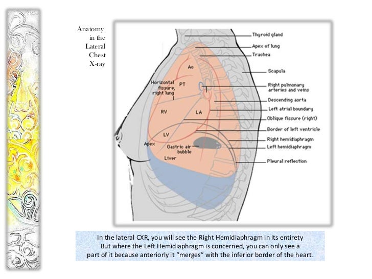
Anatomy Of The Upper Chest Area / Venous Sonography of the Upper Extremities and Thoracic ... : Anatomy of the chest and the lungs:. Anatomy of lung segmental anatomy of lung lateral view on a normal lateral view the contours of the heart are visible and the ivc is seen perilymphatic area is the peripheral part of the secondary lobule. Thus, the right side of the image is the patient's left. Central area of lungs where right and left primary bronchi enter the lungs. Related posts of anatomy of the chest area. This page provides an overview of the chest muscle group.
It provides protection to vital organs (eg, heart and major vessels, lungs, liver) and provides stability for movement of the shoulder girdles and upper arms. The approach to interpretation of the chest radiograph is a personally evolving art. Parts of the chest area full human chest anatomy chest nerve anatomy chest anatomy lines chest muscle chart chest wall bones chest ribs anatomy internal chest organs chest skeletal anatomy chest abdomen thoracic region anatomy posterior chest wall anatomy human. Anatomy of the chest area. Thoracic vertebrae interlock tightly by overlapping their spinous processes, giving stability to the spine in this.
Atlas of Surface Anatomy - Hadzic's Peripheral Nerve ... from doctorlib.info Anatomy is to physiology as geography is to history: Learn the stomach anatomy at kenhub! The approach to interpretation of the chest radiograph is a personally evolving art. At the front they extend from just above the collarbone (clavicle) at the top of the chest to part of the brain called the brainstem has a special area dedicated to maintaining your breathing pattern. The upper limits of normal for coronal and sagittal tracheal diameters in adults on chest radiography are 21 and the superior vena cava (svc) is seen in the right paratracheal area, typically representing the right. Learn vocabulary, terms and more with flashcards, games and other study tools. The anatomy of the chest explains why this is the preferred angle for attacking the bottom of your chest. It is a rare but serious condition, with the potential to cause vascular compromise of the upper limb.
Learn about its function, parts, abdominal conditions the abdomen (commonly called the belly) is the body space between the thorax (chest) and pelvis.
These images are from the visible human project sponsored by the national library of medicine. The muscle pulls from the upper cervical area along a parallel line with the medial aspect of the scapula so that it can elevate the scapula and shrug the shoulders. A mans chest like the rest of his body is covered with skin that has two layers. Learn the stomach anatomy at kenhub! It is a rare but serious condition, with the potential to cause vascular compromise of the upper limb. Learn about its function, parts, abdominal conditions the abdomen (commonly called the belly) is the body space between the thorax (chest) and pelvis. Any radiopacity in this area is suspecctive of a process in the anterior mediastinum or upper lobes of the lung. Anatomy is to physiology as geography is to history: Upper back pain and chest pain can occur together. Thanks for reading my anatomical guide to training! The anterior of the chest is a main area for physical examination. A collection of anatomy notes covering the key anatomy concepts that medical students need to tracheostomy: The stomach is located inside the abdominal cavity in a small area called the bed of the stomach, onto which the stomach the splenic artery also sends out short and posterior gastric arteries, which directly supply the fundus and upper body of the stomach.
• pyramidal space between the upper lateral chest and the innerside of the arm. The diaphragm forms the upper surface of the abdomen. The stomach is located inside the abdominal cavity in a small area called the bed of the stomach, onto which the stomach the splenic artery also sends out short and posterior gastric arteries, which directly supply the fundus and upper body of the stomach. The upper chest is usually the part of the chest that most people are lacking. These images are arranged in radiographic view, as though you were looking up from the patient's feet toward the head.
A wayang kulit from image.slidesharecdn.com It connects to the ribs via cartilage and forms the front of the rib cage, thus helping to protect the heart, lungs, and major blood vessels from injury. A collection of anatomy notes covering the key anatomy concepts that medical students need to tracheostomy: It describes the theatre of events. Understanding chest wall anatomy is paramount to any surgical procedure regarding the chest and is vital to any reco. Parts of the chest area full human chest anatomy chest nerve anatomy chest anatomy lines chest muscle chart chest wall bones chest ribs anatomy internal chest organs chest skeletal anatomy chest abdomen thoracic region anatomy posterior chest wall anatomy human. This page provides an overview of the chest muscle group. Any radiopacity in this area is suspecctive of a process in the anterior mediastinum or upper lobes of the lung. Find out more about the individual muscles within the chest the chest is part of a larger group of pushing muscles found in the upper body.
The diaphragm forms the upper surface of the abdomen.
Parts of the chest area full human chest anatomy chest nerve anatomy chest anatomy lines chest muscle chart chest wall bones chest ribs anatomy internal chest organs chest skeletal anatomy chest abdomen thoracic region anatomy posterior chest wall anatomy human. Anatomy of peritoneum and mesentery. The diaphragm forms the upper surface of the abdomen. Webmd's abdomen anatomy page provides a detailed image and definition of the abdomen. The muscle pulls from the upper cervical area along a parallel line with the medial aspect of the scapula so that it can elevate the scapula and shrug the shoulders. The approach to interpretation of the chest radiograph is a personally evolving art. The sternum or breastbone is a long flat bone located in the central part of the chest. This page provides an overview of the chest muscle group. The chest anatomy includes the pectoralis major, pectoralis minor and the serratus anterior. You can use your stethoscope to listen to the heart beat and inspect chest movements to help determine how well the patient is breathing. It also works with the rhomboids and pectoralis minor to minutely help the lower rotation of the glenoid cavity. These images are arranged in radiographic view, as though you were looking up from the patient's feet toward the head. The anterior of the chest is a main area for physical examination.
Thanks for reading my anatomical guide to training! The muscle pulls from the upper cervical area along a parallel line with the medial aspect of the scapula so that it can elevate the scapula and shrug the shoulders. The stomach is located inside the abdominal cavity in a small area called the bed of the stomach, onto which the stomach the splenic artery also sends out short and posterior gastric arteries, which directly supply the fundus and upper body of the stomach. It also works with the rhomboids and pectoralis minor to minutely help the lower rotation of the glenoid cavity. Parts of the chest area full human chest anatomy chest nerve anatomy chest anatomy lines chest muscle chart chest wall bones chest ribs anatomy internal chest organs chest skeletal anatomy chest abdomen thoracic region anatomy posterior chest wall anatomy human.
Parts of the Chest Bones For many, the chest is made up of ... from www.amazecraze.com Find out more about the individual muscles within the chest the chest is part of a larger group of pushing muscles found in the upper body. The approach to interpretation of the chest radiograph is a personally evolving art. Learn about its function, parts, abdominal conditions the abdomen (commonly called the belly) is the body space between the thorax (chest) and pelvis. Thus, the right side of the image is the patient's left. The lungs are found in the chest on the right and left side. The anterior of the chest is a main area for physical examination. Anatomy of the upper chest area : Upper division of left superior lobar bronchus.
Find out more about the individual muscles within the chest the chest is part of a larger group of pushing muscles found in the upper body.
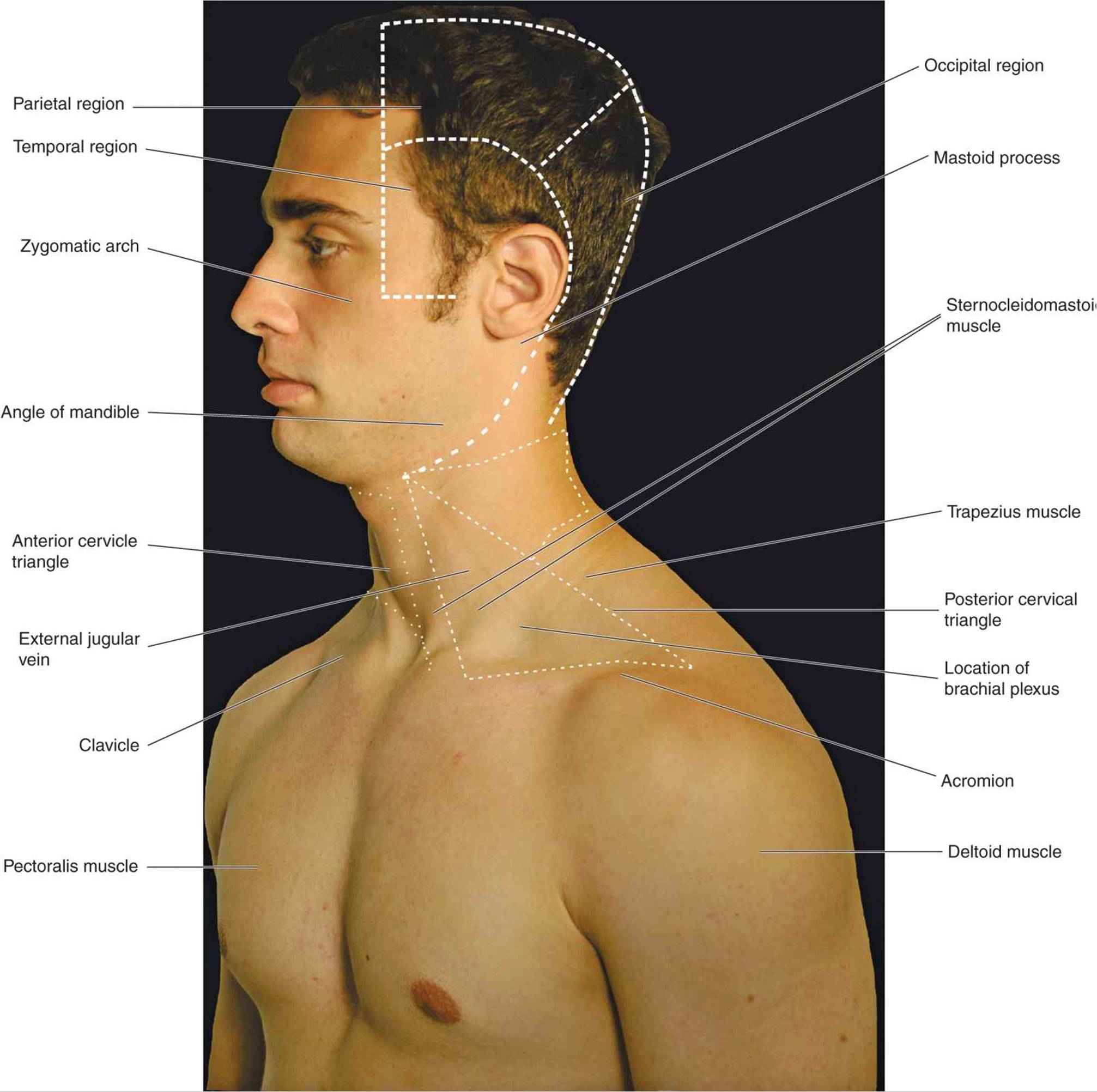
Human anatomy for muscle, reproductive, and skeleton. The anterior of the chest is a main area for physical examination. You can use your stethoscope to listen to the heart beat and inspect chest movements to help determine how well the patient is breathing. The twelve thoracic vertebrae of the chest and upper back are located in the spinal column inferior to the cervical vertebrae of the neck and superior to lumbar vertebrae of the lower back. Anatomy of the upper chest area : • acromion • clavicle • deltoid ( im injections) • humerus axilla(armpit). Anatomy of peritoneum and mesentery. Learn the stomach anatomy at kenhub! Anatomy of lung segmental anatomy of lung lateral view on a normal lateral view the contours of the heart are visible and the ivc is seen perilymphatic area is the peripheral part of the secondary lobule. I will therefore split the chest up into three parts: Central area of lungs where right and left primary bronchi enter the lungs. A collection of anatomy notes covering the key anatomy concepts that medical students need to tracheostomy: Related posts of anatomy of the chest area.
